Quizlet is the easiest way to study, practice and master what you’re learning. [1, 2] the pr interval of the surface ecg is measured from the onset of atrial depolarization (p wave) to the beginning of ventricular depolarization (qrs complex).

Lyme disease) electrolyte disturbances (e.g.
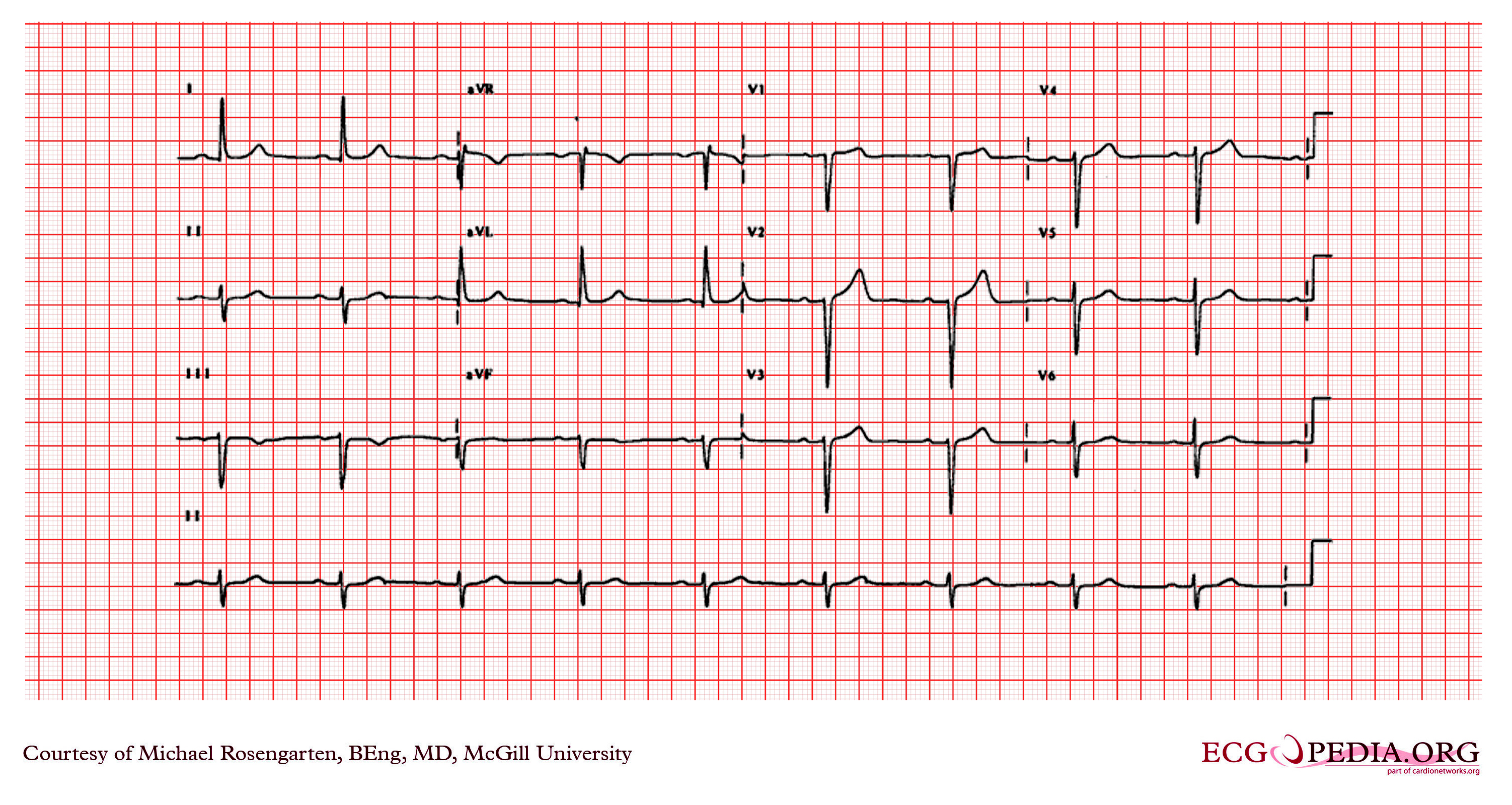
First degree heart block strip. First degree av block is a rhythm that is not actually a real block, but more of a slowing down. P waves are present and lengthening, the qrs complexes are 0.10 seconds, and the ventricular rate is irregular but 70 beats per minute. This condition is generally asymptomatic and discovered only on routine ecg.
The atria contract and then there is an abnormal delay before the ventricles contract. It usually does not cause problems. Usually the delay occurs in the av node.
It is cause by a conduction delay at the av node or bundle of his. Try to think of it like traffic, when a freeway closes a lane and traffic is funneled through a smaller. Heart blocks are arrhythmias caused by conduction disturbances at the av node.
More than 50 million students study for free with the quizlet app each month. Telemetry interpretation is an important skill for nurses in many patient care areas. Create your own flashcards or choose from millions created by other students.
[1, 2] the pr interval of the surface ecg is measured from the onset of atrial depolarization (p wave) to the beginning of ventricular depolarization (qrs complex). You begin your shift and assess an electrocardiogram rhythm strip. Failure of some of the p waves to propagate into the ventricles.
As the signal travels from the sa node to the av node and on through the junction, it gets slowed down. 3rd degree heart block happens when the impulse from the sa node is totally blocked at the av node, and nothing passes through to the ventricles. This means than the pr interval will be longer than normal (over 0.20 sec.).
The overall heart rate is 30 beats per minute. There is no electrical block but rather a slowing or delay of the signal. After watching the cardiac monitor, you notice the rhythm has suddenly changed.
Lyme disease) electrolyte disturbances (e.g. Treatment of a 1st degree av block is usually not necessary, however, it�s important to monitor the patient�s rhythm to make sure it doesn�t progress into a more severe block. First degree heart block is actually a delay rather than a block.
159 sinus rhythm with 1st degree av block (pr 0.22 seconds) and pacs. Describe the basic electrophysiology of the heart. Second degree av block type i and 2:1 av block ecg 2:1 av block ecg (example 1) 2:1 av block ecg (example 2) 2:1.
A first degree av block causes a prolonged impulse conduction time from the atria to the ventricles, due to a delay in the av node. Normally, this interval should be. Identify basic components of an ecg strip.
In addition, this is what will be present with a 1 st degree av heart block: This slight difference in nomenclature is usually overlooked by many. Causes of first degree heart block.
Enlarge recall that the p wave indicates atrial depolarization, initiated by firing of. It is defined by ecg changes that include a pr interval of greater than 0.20 without disruption of atrial to ventricular conduction. This is commonly seen in athletes.
Note that this is still a sinus rhythm, but not normal sinus rhythm. Second degree heart block type ii. The qrs complexes are 0.18 seconds.
The p waves are absent and qrs complexes are regular. The pr interval is greater than 0.2 seconds long. Quizlet is the easiest way to study, practice and master what you’re learning.
This lengthening of the pr interval is caused by a delay in the electrical impulse from the atria to the ventricles through the av node. First degree heart block will look like a typical sinus rhythm with one distinguishing feature. Rhythm strip flash card practice.
Distinguish between the degrees of heart blocks by identifying rhythm strips. The rate will be that of the underlying rhythm. Atrioventricular blocks (otherwise known as av blocks or heart blocks) can be divided into three degrees.
120 3rd degree heart block (complete heart block) 121 2nd degree av block mobitz ii (2 or more p waves for every qrs) 122. Describe the causes of atrioventricular heart blocks. The electrical impulse flowing through the heart is delayed when moving from the atria to the ventricles in a first degree heart block.
If you don’t know how to measure a pr interval see this article and video. Rate, regularity, p wave morphology and qrs duration and morphology will be unaffected. The nurse assesses the cardiac rhythm as: