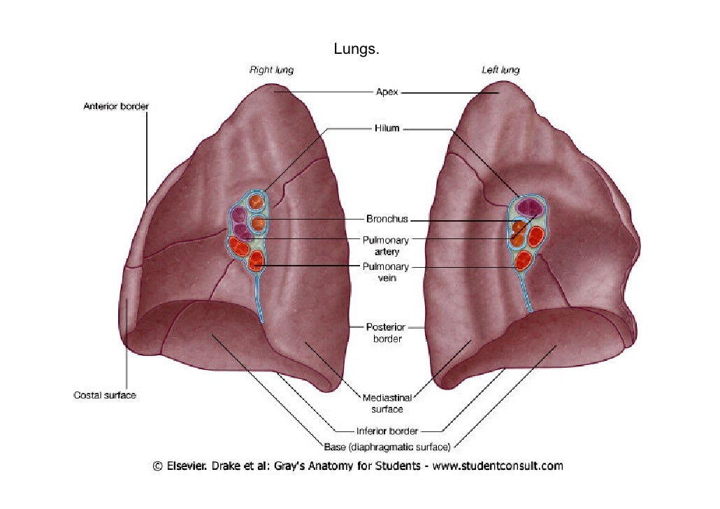
The hilum is an area while the root of the lung is the structures that enter and exit through the hilum. What does the medical term hilar mean?

Of, relating to, affecting, or located near a hilum hilar lymph nodes of the lung.
Right hilum of lung. A prominent right hilum is an enlargement of the root of the lung. The lung hilum (where structures enter and leave the lung) is located on this surface. Hilum and root of the lung are two components of the lungs.
This region aids the lung�s root. Both human lungs have a hilar region, meaning both lungs have an area called the hilum. This may be a normal variation or consequent to a disease process.
It rests on the dome of the diaphragm, and has a concave shape. Of, relating to, affecting, or located near a hilum hilar lymph nodes of the lung. The hilum is what connects your lungs to their supporting structures and where pulmonary vessels enter and exit your lungs.
Anatomy of the hilum both the right and the left lung have a hilum which lies roughly midway down the lungs, and slightly towards the back (closer to the vertebrae than to the front of the chest). Hilum of the right lung. Medical experts tend to divide the hilum of the lung into “left hilum,” and “right hilum.” the left hilum of the lung is large, dense and pulled laterally and upwards to the left.
The hilum of the lung is found on the medial aspect of each lung, and it is the only site of entrance or exit of structures associated with the lungs.that is to say, both lungs have a region called the hilum, which serves as the point of. On chest radiographs, the lung hila have a. The right and the left root of the lung is not identical due to the pulmonary artery.
Missing right breast shadow (mastectomy) clinical information. The right hilum is bigger than the left; This can be sorted out in the perspective of the clinical picture (symptoms and signs), appropriate laboratory tests and if necessary a ct scan.
He hilum of the right lung is arched by azygous vein. The hilum is a large triangular depressed area, or a fissure, on the lung located on the medial aspect of each lung. Both are on the medial surface of the lung.
The left upper lobe (lul) and the left lower lobe (lll). Right and left lung are separated by the mediastinum. 1.2).the root of the right lung lies behind the svc, the superior part of the right atrium, and below the azygos vein.
Uncovering the underlying cause of a prominent right hilum may require the use of. Thus, the upper lobe bronchus and artery are found above the level of the right. The dense hilum sign suggests a pathological process at the hilum or in the lung anterior or posterior to the hilum.
The right and left lung anatomy are similar but asymmetrical. The hilar region is where the bronchi, arteries, veins, and nerves enter and exit the lungs. The structures within the right hilum are arranged such that the principal bronchus.
The base of the lung is formed by the diaphragmatic surface. Pulmonary artery is the uppermost structure in left lung hilum. Malignancy, especially lung cancer, should be suspected.
Each lung has a hilum and root of the lung. The hilum is an area while the root of the lung is the structures that enter and exit through the hilum. The right lung consists of three lobes:
The left lung consists of two lobes: The right lobe is divided by an oblique and horizontal fissure, where the horizontal fissure. A hilum is a section of an organ where other types of structures like veins or arteries can enter.
In the right hilum the bronchus of the upper lobe and the branch of the right pulmonary artery to the upper lobe originate prior to entering the hilum. Often, hilar enlargement is due to enlargement of these nodes. The right upper lobe (rul), the right middle lobe (rml), and the right lower lobe (rll).
Posterior relation of hilum of lung is vagus nerve. What does the medical term hilar mean? Anatomically, the hilum of an organ is the point of entry of the neurovascular bundle (arteries, veins and nerves) into an organ.
The hilum — or root —. In addition, the lymphatics and lymph nodes are typically along for the ride. The hilum of the lung (also called the root of the lung) is formed by the principal bronchi, the central pulmonary arteries and veins, the bronchial nerves and vessels, and the lymphatics, which enter and leave the lung from the mediastinum ( fig.
Passes inferolaterally, inferior to the arch of the aorta and anterior to the esophagus and thoracic aorta, to reach the hilum of the lung. The right hilum is caudally related to the terminal azygos vein and posteriorly related to the right atrium and superior vena cava. A prominent hilum refers to an apparent enlargement of the root of the lung.
Gross anatomy of lungs lungs are a pair of respiratory organs situated in a thoracic cavity. It may exist naturally due to normal variation of the structure, or it may be caused by a disease. This concavity is deeper in the right lung, due to the higher position of the right dome overlying the liver.
Hilum of lung in this image, you will find a root, hilum, bronchus, pulmonary artery, deoxygenated blood, pulmonary vein, oxygenated blood, pulmonary ligament, right lung, left lung in it.