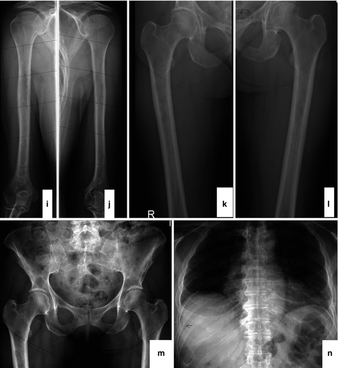
Loss or thinning of bone (osteoporosis or osteopenia) holes in bone (lytic lesions), and/or. Skeletal survey in advanced multiple myeloma:

Traditionally, evaluation for destructive bone lesions from multiple myeloma has been performed using a skeletal survey employing multiple plain radiographs of a patient.
Skeletal survey for multiple myeloma. How to better afford the high cost of myeloma therapy? To compare the evaluation of myeloma bone involvement by conventional radiographic skeletal survey (rss) with whole body magnetic resonance imaging (mri). In an attempt to compare the sensitivity of bone radiographs and bone marrow magnetic resonance (mr) imaging for bone lesion detection in patients with stage iii multiple myeloma (mm) and to evaluate the possible consequences of the replacement of the conventional.
Per nccn guidelines for smoldering multiple myeloma (smm), whole body radiography, i.e. With these results, phan wanted to do a full skeletal survey to see if there are any identifiable weaknesses or holes in my bones.and to make sure i have no lesions. For decades, conventional skeletal survey (css) has been the standard imaging technique for multiple myeloma (mm).
The skeletal survey is used at the time of diagnosis for the purposes of �staging� the disease. So the 6 and half years of steroids is the culprit for the weakening bones. This analysis compares sensitivity and prognostic significance of wbct and css in patients with smoldering mm.
A s keletal survey demonstrates innumerable well circumscribed �punched out� lytic lesions best seen in the lateral view of the skull. Loss or thinning of bone (osteoporosis or osteopenia) holes in bone (lytic lesions), and/or. A study of the international myeloma working group.
For decades, conventional skeletal survey (css) has been the standard imaging technique for multiple myeloma (mm). This is called a skeletal survey. Bone disease is the hallmark of multiple myeloma.
Due to this, imaging procedures play a key role in the management of multiple myeloma. Radiographic versus mr imaging survey. Due to this, imaging procedures play a key role in the management of multiple myeloma.
Skeletal surveys in multiple myeloma skeletal surveys in multiple myeloma sebes, j.; Multiple myeloma, characterized by the clonal proliferation of plasma cells in the bone marrow, is the second most common hematologic malignancy, with more than 20,000 new cases diagnosed per year in the united states (laubach, richardson, & anderson, 2010).typical clinical features include anemia, renal failure, hypercalcemia, and skeletal lytic lesions (kyle & rajkumar, 2004). Skeletal survey in advanced multiple myeloma:
Skeletal survey, with its low sensitivity, does not rise to the challenge. In myeloma, the large number of plasma cells being made in the bone marrow can cause damage to the hard outer covering of the bones. Wb radiography has been the reference standard method for mm evaluation for the past decade.
19 of the patients had newly diagnosed multiple. Skeletal lesions are evaluated to establish the diagnosis, to choose the therapies and also to assess the response to treatments. A skeletal survey is essential in patients suspected of myeloma.
Twenty receiving chemotherapy were also followed with skeletal surveys in order to evaluate bone response to treatment. Skeletal survey is characterized by wb radiography, including imaging of the cervical, thoracic, and lumbar spine; A close association was found between skeletal findings and.
Bone disease is the hallmark of multiple myeloma. The crab disease spectrum and myeloma biomarkers must be absent. I think that if lesions can be seen, the myeloma is then considered to be stage 1 or higher, compared to being classified as smm.
For decades, conventional radiography has been the standard imaging modality. The study found the skeletal survey had a sensitivity of just 57.1% and a specificity of 86.9%. Skeletal survey (ss), should be used to rule out osteolytic bone lesions.
A study of the international myeloma working group. Because smoldering multiple myeloma patients typically have a low burden of disease, ruling out bone disease requires a highly sensitive imaging modality, the researchers said. Skeletal lesions are evaluated to establish the diagnosis, to choose the therapies and also to assess the response to treatments.
Traditionally, evaluation for destructive bone lesions from multiple myeloma has been performed using a skeletal survey employing multiple plain radiographs of a patient. Hillengass j, moulopoulos la, delorme s, et al. • in marked radiographic osteopenia and obesity, ldb ss diagnostic performance is reduced.
Skeletal survey in multiple myeloma: Features are in keeping with multiple myeloma. My myeloma is under control and i haven�t had any bone involvement from the disease.