In confocal microscopy, the use of more than 256 gray levels is seldom justified by the detection resolution, although there may be some value in discriminating more intensity levels for certain data processing algorithms. The confocal optics provides a unique characteristic that a conventional microscope lacks.

In an ordinary simple microscope, light passes through the sample, whereas in a confocal microscope focuses a smaller beam of light at one narrow depth level at a time.
What is confocal microscopy. It is important to recognize that pixelation at the image display stage is not unique to digital imaging. To achieve this task, the sample is scanned by a tiny light point. Confocal microscopy can be used to create 3d images of the structures within cells.
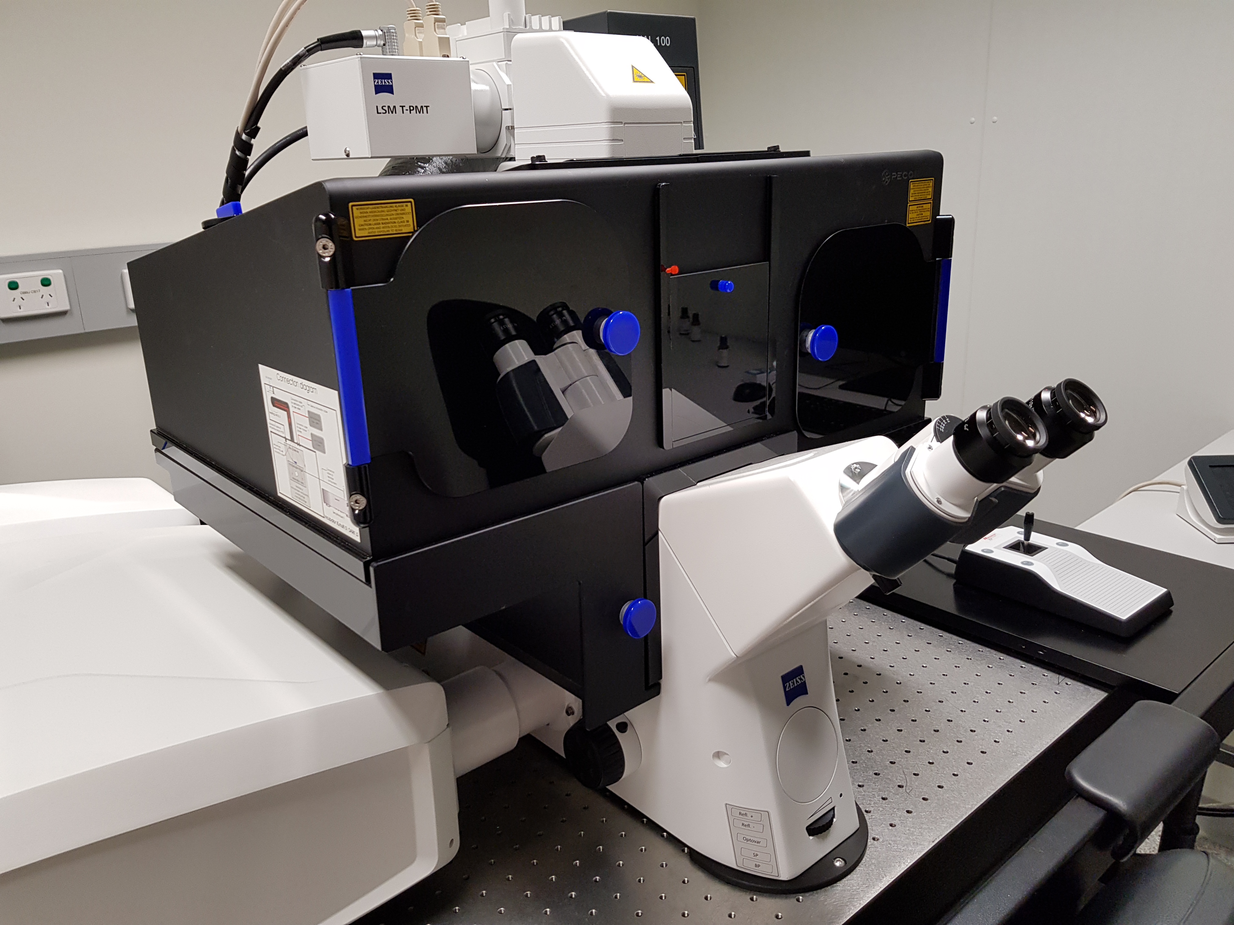
In an ordinary simple microscope, light passes through the sample, whereas in a confocal microscope focuses a smaller beam of light at one narrow depth level at a time. Neurons and astrocytes population labeled with different fluorophores. The confocal microscope attachment shown in these pictures contains the optics for scanning the laser beam, and the pinhole.
The confocal also includes a very large box containing electronics, which is not shown in the photographs; Confocal microscopy is broadly used to resolve the detailed structure of specific objects within the cell. Confocal microscopy is a major advance upon normal light microscopy, allowing one to visualise not only deep into cells and tissues, but also to create three dimensional images and to follow specific cellular reactions over extended periods of time.
Examining these structures can help researchers observe the internal workings of cellular processes. Confocal microscopy is a powerful technique to image fluorescent samples given its high efficiency, low background noise and the capability to optically section samples into different focal planes. Optical sectioning is a common application in the biomedical sciences and has been useful for.
The molecules, called fluorophores, release photons as they return to their unexcited state, causing the fluorescence. Confocal microscopy is a specific form of fluorescence microscopy wherein a sample is. The system is composed of a a regular ßorescence microscope and the confocal part, including scan head, laser optics, computer.
On images captured using confocal options, the areas in focus will be highlighted. Optimally, the slices cover just the depth of focus generated in the microscope. In confocal microscopy, the use of more than 256 gray levels is seldom justified by the detection resolution, although there may be some value in discriminating more intensity levels for certain data processing algorithms.
Confocal microscopy (hamilton and wilson, 1982) is widely used, not only for fluorescence microscopy and 3d sectioning of transparent materials, but for the measurement of surface topography when used in reflection mode. There is also a laser, and an sgi computer. A standards document, which describes confocal microscopy and its influence quantities, has recently.
A focused laser beam is used to excite the molecules at one point of the sample. Hi #measuringhero!let�s learn how confocal microscopy works and how i solved a metrology challenge with that.learn more about #measuringhero: Example of a confocal microscopy micrograph.
Confocal fluorescence microscopy is a commonly used optical imaging method in biology, combining fluorescence imaging with confocal microscopy for. Confocal microscopy offers several advantages over conventional widefield optical microscopy, including the ability to control depth of field, elimination or reduction of background information away from the focal plane (that leads to image degradation), and the capability to collect serial optical sections from thick specimens. Fluorescence microscopy is a general category of imaging where a sample is labelled with “glowing” molecules that make it easier to see;
In conventional widefield optical microscopes, secondary fluorescence emitted from a specimen often occurs through the excited volume and obscures resolution of. The confocal optics provides a unique characteristic that a conventional microscope lacks. As a distinctive feature, confocal microscopy.
The smallest shape that can be generated is a diffraction limited spot. Hi dayan, if you have an unused inverted microscope frame available somewhere in your department, it might be worth looking the confocal.nl. Similar to widefield fluorescence microscopy, various components of living and fixed cells or tissue sections can be specifically labeled using immunofluorescence, for example, and then visualized in high resolution.
It is known as optical sectioning. The basic key to the confocal approach is the use of. Confocal microscopes allow to optically separate slices of the sample’s response and record theses slices as images.
Spinning disk, also known as the nipkow disk, is a type of confocal microscope that uses several movable apertures (pinholes).